This Computer Vision solution is related to medicine. The client’s company works to discover a cure for Alzheimer and other neurodegenerative diseases. It is their ambitious research, which is striving for a ground-breaking approach in the drug discovery. To test whether the medicine works or not, it is injected in the cells. Then the scientists are checking these cells through the microscope, to see if they are still alive or not, they do it once a day over 10 days. The project deals with determining the lifetime of each cell. The scientists have to remember that the cells are sensitive to the light; therefore, they are watched through the microscope once a day.
There are three main project objectives:
The main challenge on the project was to separate one cell from another. To be more specific: the cells are placed in a Petri dish and can form groups. So, we need to detect and separate each cell in the group to determine whether it’s alive or not.
To solve this problem, our developer used mathematical methods such as graph theory methods.
So, here is how it works. The end solution takes a picture as an input. For the first step, we have to detect the ‘single’ little cells on the original picture. Then the picture is blurring and we’re trying to detect big cells and cell groups. After that, we remove these big cells and try to separate each cell from the others in the group.
When all of the cells are found, we have to determine whether they still have neurites. Neurites are processes of the cell’s nerves. The presence of the neurites allows us to determine whether the cell is alive or not.
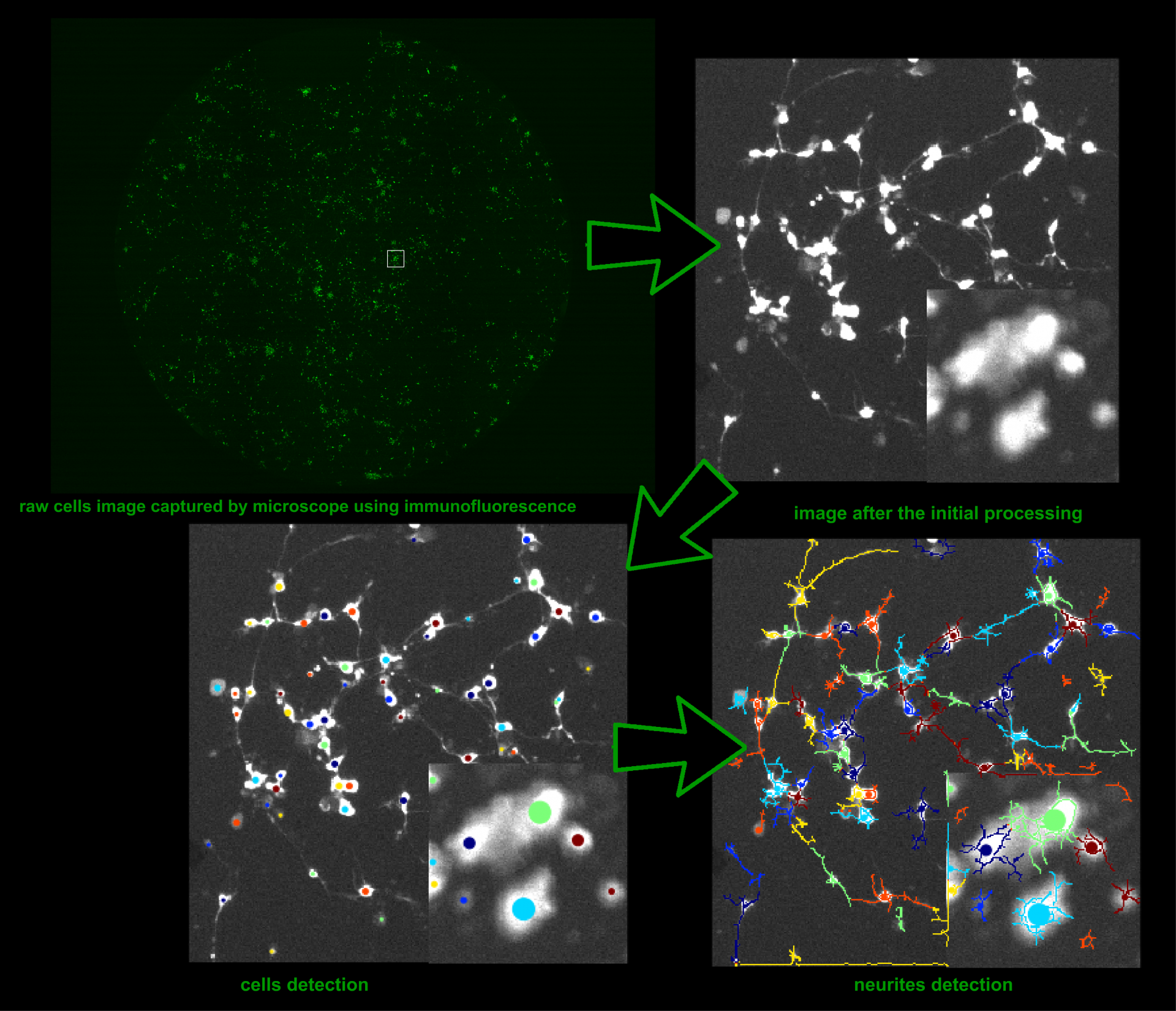
As a result of the project, the script was developed. As an input, it takes 10 images made via the microscope. Then the script recognizes the live and dead cells and marks them on the pictures. As an output, the script returns the images with the marked cells and several text reports which contain detailed information about the cells. The script is currently being used by our customer and is already helping them in their work.